For my own personal use only:
-
Columnaris disease
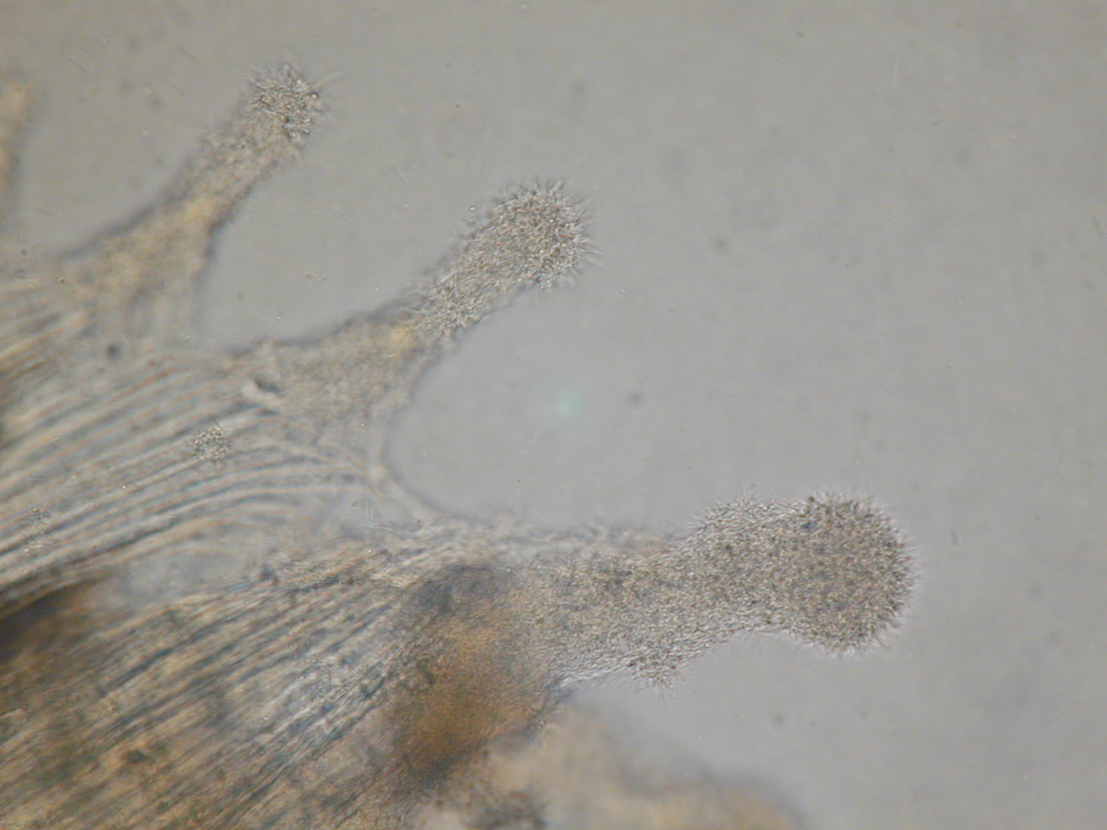
High magnification photomicrograph of a fin biopsy showing a clump of Flavobacterium columnare bacteria – note the commonly seen “haystack” formations on the edges of fin rays -
Classic case:
- “Saddleback” lesions are patches of pale discoloration of skin +/- red edges located dorsally (at the base of and around the dorsal fin)
- Gills may have patchy white or tan discolored areas (i.e., “necrosis”)
- These and other affected areas of fish may have increased mucus/slimy exudate
- Fin deterioration
- Anorexia, oral mucosal erosion and/or ulceration
-
Dx:
Etiology: Flavobacterium columnare is a gram-negative, rod-shaped, long and motile filamentous bacterium
-
Dx: see “waving haystacks” of bacteria on wet mount slides of lesions from skin, fin, or gills
- Although this is considered “classic,” these are not always evident on wet mount evaluation
-
Dx: see “waving haystacks” of bacteria on wet mount slides of lesions from skin, fin, or gills
-
Tx:
- Early infection/bath treatment: add potassium permanganate, hydrogen peroxide, or diquat to the water
- Chronic disease: bath treatment as above but may also need to treat with medicated feed containing florfenicol or oxytetracycline
- Prevent: minimize traumatic injuries, reduce organic debris in the tank, including uneaten feed
- Pearls:
- Environmental or handling factors often compromise skin/mucus and predispose to columnaris infection
- Can spread rapidly
- Uneaten food may serve as a reservoir
- There is a columnaris vaccine in the U.S. for largemouth bass and channel catfish
-
Classic case:
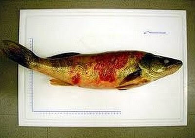
- Motile aeromonad septicemia
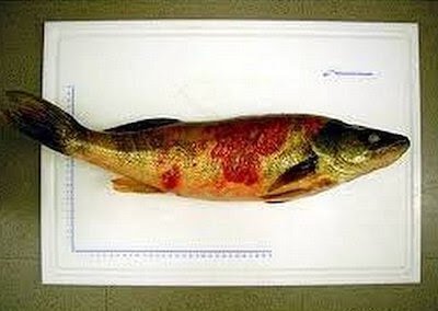
Motile aeromonad septicemia in a fish - Classic case:
- Variable morbidity and mortality rates (depend on water quality, dissolved oxygen level, stress level)
- External hemorrhages, petechiae, and/or erythema; can have ulceration
- Hemorrhages/petechiae in the viscera, body wall, and peritoneum
- Other non-specific signs may be present, including ascites
- Dx:
- Etiology: most often caused by motile Aeromonas spp. infection in freshwater fish (Vibrio spp. are more common cause in marine fish)
- Often “secondary” to poor management or husbandry issues
- Dx: bacterial culture of kidney, brain, other affected organs
- Etiology: most often caused by motile Aeromonas spp. infection in freshwater fish (Vibrio spp. are more common cause in marine fish)
- Tx:
- Correct underlying stressors including environmental or other management issues
- Antibiotics, based on culture and susceptibility
- Pearls:
- Risk factors include poor water quality (including low dissolved oxygen levels, elevated ammonia), trauma, and handling stress
-
“Red sore disease” is a common manifestation of this seen in wild game fish
- Combination of motile Aeromonas sp. infection and protist parasite infection (often Epistylis or Heteropolaria both sessile ciliates)
- Classic case:
- “Ich” (freshwater) or “white spot disease” (freshwater or marine)
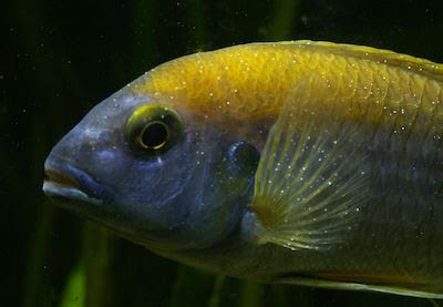
Ich in a freshwater fish; note the white spots all over the surface - Classic case:
- Small white spots all over fish (body, gills, fins)
- Extreme lethargy, inappetance, mortality
- Dx:
- Freshwater etiology: Ichthyophthirius multifiliis (“Ich”) is a ciliated obligate parasitic protozoa that can reproduce exponentially (hundreds to a thousand offspring in one life cycle)
- Marine etiology: Cryptocaryon irritans has a similar “explosive” numbers-type life cycle
- Dx: microscopic evaluation of skin, fin, and/or gill scrape or biopsy -- make a slide to see characteristic ciliated protozoa with gray/granular interior that rotates slowly or moves like an amoeba
- This life stage, known as the feeding stage or “trophont,” is often embedded under skin or mucus (when seen in tissue biopsies)
- For mature freshwater Ichthyophthirius stages, horseshoe-shaped macronucleus is more apparent
- For marine Cryptocaryon, internal structures are not easily seen in any stages.
- Tx: only free-swimming, infective stages (“theronts”) in the water for both freshwater Ichthyophthirius and marine Cryptocaryon are susceptible to treatment!
- Pet fish: multiple treatments of formalin or malachite green (the latter cannot be dispensed by vet but may be available over-the-counter)
- Alternatively, for many freshwater species, 4-5 g/L salt for an extended period of time, depending on temperature
- Copper therapy
- Refractory to treatment: parasites encysted in the environment and those on the fish
- Pet fish: multiple treatments of formalin or malachite green (the latter cannot be dispensed by vet but may be available over-the-counter)
- Pearls:
- Life cycle varies depending on temperature - at “warmer” temperatures (78–80ºF [25.5–26.6ºC]), freshwater Ichthyophthirius treatment period may last a week or more
- For Cryptocaryon in marine tropical fish, treatment duration may be 4–6 weeks because the life cycle of Cryptocaryon is significantly longer than that of freshwater Ichthyophthirius
- Recovered fish can be silent shedders (carriers) in the future and will have some degree of immunity
- The classic white spot of “Ich” seen grossly is actually the encysted parasite surrounded by tissue reaction by the fish
- In younger stages of freshwater Ichthyophthirius, the “U” or “C” shaped macronucleus may not be apparent
- Classic case:
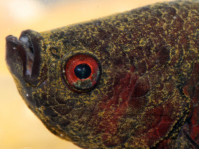
- Velvet disease (freshwater and marine)

Velvet disease in a fish - Classic case:
- Swimming against objects or substrate (flashing), lethargy, gathering near the surface more than usual
- Affected fish have a dusty, velvet appearance, but this may not be readily apparent in lighter colored fish or in predominantly gill infestations
- Dx:
- Etiology: Amyloodinium ocellatum is a parasitic dinoflagellate that attaches to, and invades, the skin of brackish to marine fish (salinity of 3–45 ppt)
-
Etiology: Piscinoodinium pillulare (and several others) are freshwater counterparts to Amyloodinium, and are very similar in appearance microscopically
- Both marine and freshwater parasites can have explosive population growth in one life cycle - i.e., one adult may lead to hundreds of organisms in one generation
- Dx: wet mount preparation of gill tissue shows many small pale brown/golden organisms (trophonts)
- Microscopic exam: round, ovoid, or pear-shaped, brown/golden trophonts (feeding stages) are evident anchored to the gill epithelium, fins, and skin
- Tx:
- Rx for pet fish can include use of a chloroquine extended bath
- Extended Rx of copper sulfate is one of the few legal options for food animals, but can also be used in marine pet fish and in freshwater (in many instances)
- A pH-adjusted freshwater dip (0 ppt, for 3–5 minutes) will help remove significant portions of the brackish to marine Amyloodinium “feeding” or trophont stage
- Some euryhaline species (those that have a wide salinity tolerance) can handle extended freshwater exposure for 2–3 weeks
- A saltwater dip (30 ppt for 3–5 minutes) will help remove the freshwater parasite in freshwater species
- Note: extended treatments which include the environment are required due to more resistant environment life stages found off the fish in both freshwater and marine infections
- Pearls:
- The feeding stage (trophont) of Amyloodinium is non-flagellated, nonmotile, and pigmented while the infective stage (dinospore) is motile
- Tomonts (the reproductive stage found off the fish and in the environment) are more resistant to treatment
- Classic case:
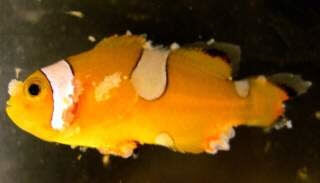
- Lymphocystis

clownfish with severe lymphocystis disease caused by Lymphocystivirus; note nodules/wart-like lesions on fins, head, and body and the erosion and ulceration on the body 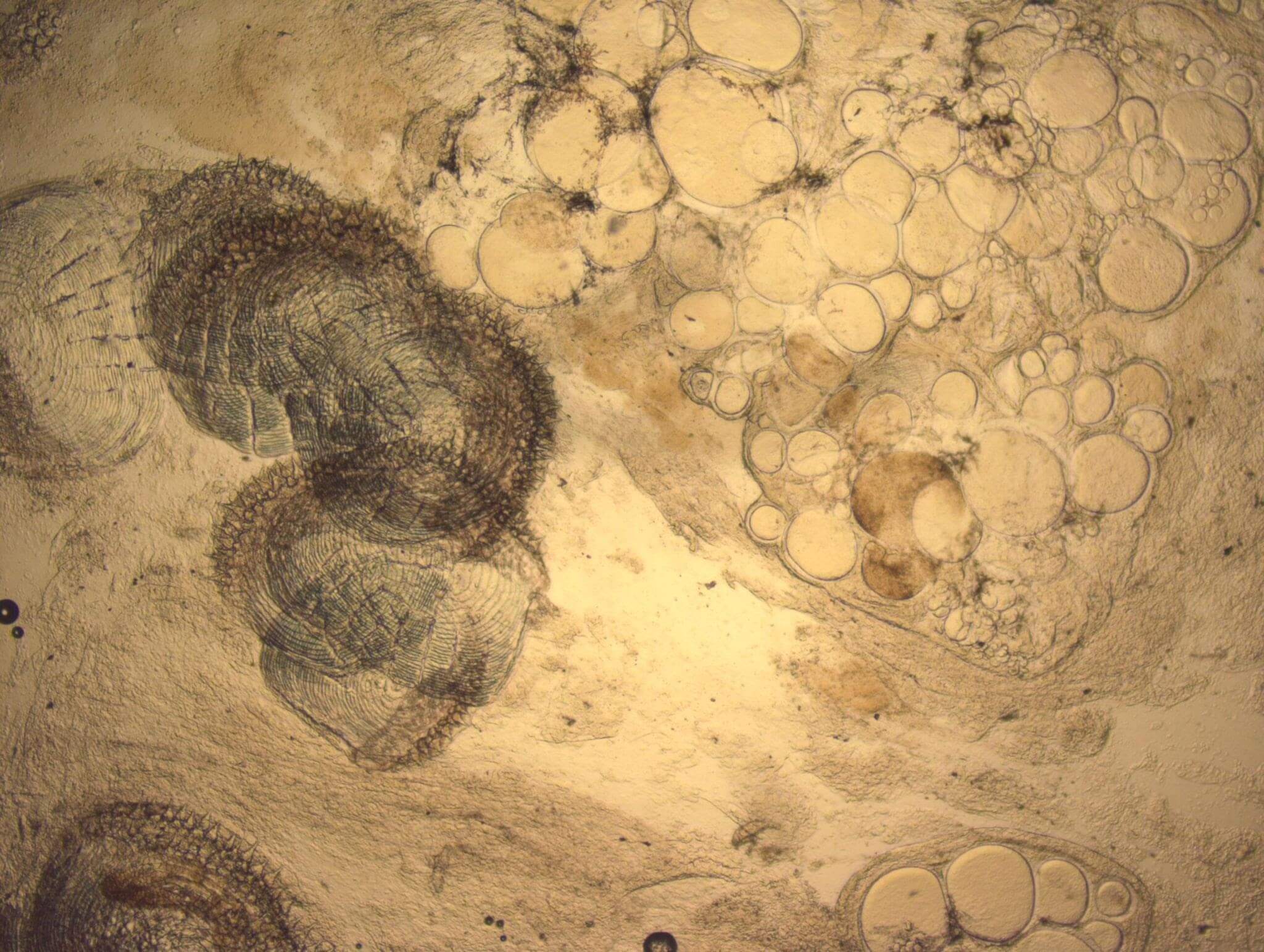
Photomicrograph of skin scrape (wet mount) from a clownfish with lymphocystis disease; note round to ovoid /balloon-shaped structures to the right of the scales - these are greatly enlarged fibroblasts filled with viral particles - Classic case:
- Papillomatous or pebble-like masses (or “warts”) on the fins, gills, or skin
- Dx:
- Etiology: infection with an iridovirus known as Lymphocystivirus or Lymphocystis disease virus (LCDV), a member of the family Iridoviridae
- Dx: microscopic evaluation of the masses reveals enlarged fibroblasts (they become “virus-making factories”) which look like “balloons”
- Tx:
- None, self-limiting in days to weeks if infection is not too severe (up to 6 weeks)
- Isolation of grossly/visibly infected specimens may help reduce viral loading and spread in the system
- Supportive care (e.g., increased or decreased salinity to aid in osmoregulation, antibiotics if secondary infections seen) may be warranted if severe infection
- Pearls:
- Mainly of aesthetic concern in “light cases”
- If significant portions of the skin or gills are affected (i.e., in moderate to more severe cases), mortalities may result from impaired osmoregulation, secondary bacterial infections, and other opportunistic diseases
- Because fibroblasts are a common cell type found in multiple organ systems, internal organs may also be affected with lesions only apparent at necropsy
- Does not affect cyprinids (e.g., koi and goldfish), catfish, or salmonids
- Contagious, but more likely if there are skin wounds/trauma or other significant stressors
- Classic case:
- Bonus! 6. Head and lateral line erosion (HLLE) disease/syndrome
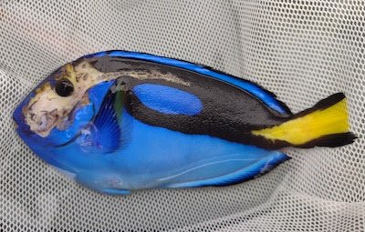
Head and lateral line erosion in a Pacific blue tang - Classic case: a.k.a. “Hole in the head disease”
- Seen in freshwater (especially in cichlids) and ornamental marine species (especially in surgeonfishes, tangs, and angelfishes)
- Erosions around eyes/on the head and along the lateral line
- Dx:
- Etiology: considered to be a syndrome, use of activated carbon has been proven as one cause in published marine studies
- Other etiologies and/or predisposing factors that are suspected but as yet unproven include nutrition, water quality, high oxidation/reduction potential (ORP), and/or infectious agents (e.g., internal parasites such as flagellates)
- Dx is based on clinical signs
- Tx:
- Improve water quality, remove activated carbon filters, address any stray voltage near tank, enhance diet/ micronutrients (including vitamins and antioxidants)
- Metronidazole if flagellate infestation is suspected or diagnosed: administer in feed or add to water
- Pearls: common!
- Underlying causes are as yet to be determined; marine and freshwater etiologies may differ
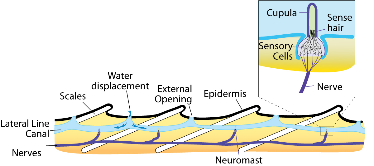
- The lateral line is part of the nervous system and helps fish navigate: helps to detect movement, vibration, and pressure differences in the water
- Click here to see a diagram of the lateral line sensory organ with neuromasts
- Fish with advanced HLLE may have irreparable scarring of skin tissue
- Classic case: a.k.a. “Hole in the head disease”


Images courtesy of Liz WinfreyV (fish tank), Roy Yanong (clownfish, columnaris, head and lateral line disease, lymphocystis, HLLE), Sneha srivatsa (hemorrhagic septicemia), djpalme (ich), mydigitallife (velvet infection), Nick Hobgood (lymphocystis).
Top Topic Category
Exotics