For my own personal use only:
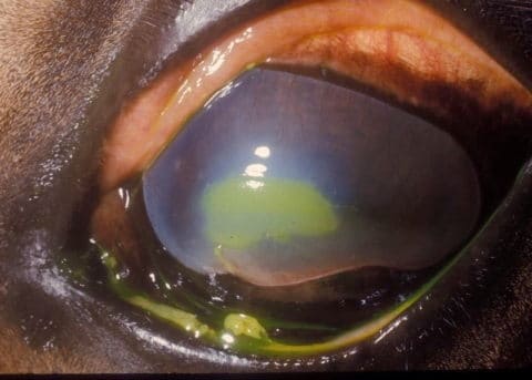
- Corneal ulcers

Superficial corneal ulcer stained with fluorescein; focal edema, miosis - Classic case: Acute onset of unilateral blepharospasm, photophobia, miosis, epiphora, corneal edema
- Dx: Thorough ophthalmic exam with ophthalmoscope, fluorescein stain positive
- Tx:
- Topical antimicrobials, atropine (mydriatic to decrease iridocyclospasm and improve drainage), and anticollagenases (e.g., serum, EDTA), +/- antifungals
- Systemic nonsteroidal anti-inflammatories
- Use subpalpebral lavage system if patient is difficult or ulcer is severe
- Surgical - conjunctival grafts for severe cases
- Pearls:
- NEVER use steroids when an ulcer is present
- Main differential is recurrent uveitis – has similar clinical signs but NO fluorescein uptake. Rx is topical steroids
- Desmetoceles and stromal abscesses are sequelae to corneal ulcers that are CRITICAL but have no stain uptake because all endothelium is gone or covered over, respectively
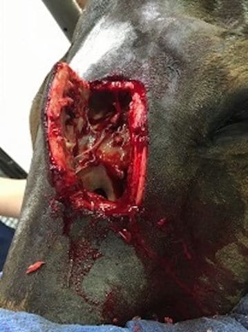
- Sinusitis

Frontal sinus flap revealing sinus cyst - Classic case: Mucopurulent unilateral nasal discharge +/- facial swelling and epiphora; often malodorous
- Dx:
- Radiographs to identify sinus or tooth pathology
- Upper airway endoscopy to evaluate drainage angles and rule out other causes of discharge
- Thorough dental examination
- Tx: Sinus trephination/flap and lavage +/- removal of mass or offending infected tooth; long-term antimicrobials
- Pearls:
- Primary - due to upper respiratory infection
- Secondary (more common) – due to dental disease, sinus cyst, ethmoid hematoma, or neoplasia
- Chronic has guarded prognosis for resolution
- Pituitary pars intermedia dysfunction (PPID, a.k.a. Cushing’s
disease)
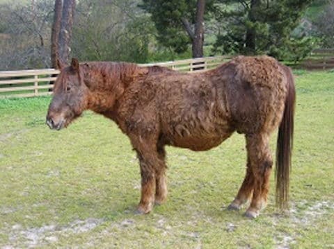
Severe hypertrichosis and muscle loss with PPID - Classic case: Horse or pony over 15 years old with chronic laminitis, hypertrichosis (long curly haircoat), recurrent infections (hoof abscesses, sinusitis), loss of topline musculature, lethargy, abnormal fat deposition (e.g., supraorbital fat pads), and polyuria/polydipsia/ polyphagia
- Dx:
- Increased resting plasma ACTH level
- Positive thyrotropin-releasing hormone stimulation test (more sensitive)
- Measure fasting insulin or do insulin sensitivity testing because most horses with PPID also have insulin dysregulation
- Tx: Daily pergolide (a dopamine agonist); have to increase dose over time as disease progresses
- Pearls:
- Lack of dopaminergic inhibition of the pituitary pars intermedia by hypothalamus leads to development of functional adenoma in pituitary pars intermedia. See increased ACTH, alpha-MSH, beta-endorphin, and cortisol
- Younger horses with regional adiposity, laminitis, and insulin dysregulation considered to have “equine metabolic syndrome”
- Colitis
- Classic case: Depression, inappetance, variable colic, decreased or hypermotile GI sounds, fever, variable degrees of shock/hypoperfusion, +/- watery or hemorrhagic diarrhea
- Dx:
- Fecal PCR panel (for Salmonella, Clostridium perfringens, C. difficile, Potomac horse fever [PHF], coronavirus), fecal egg count (for cyathastomiasis)
- Abdominal ultrasound to assess colon wall thickness (esp. right dorsal colon for NSAID-associated)
- +/- Abdominal radiographs to look for sand
- Routine labwork shows dehydration, abnormal electrolytes, WBC count (usually neutropenic), protein levels (usually hypoalbuminemic)
- Tx: Biosecurity and ...
- Supportive care – IV fluids and electrolytes and colloids
- Anti-endotoxics/anti-inflammatories (e.g., flunixin meglumine, pentoxifylline, polymyxin B, hyperimmune plasma)
- Antidiarrheals (e.g., bismuth subsalicylate, Biosponge)
- +/- Antimicrobials (metronidazole for clostridiosis, oxytetracycline for PHF, otherwise controversial)
- Put feet in ice-water slurry to prevent laminitis
- Pearls:
- Can be mild or severe and life-threatening with huge costs
- Salmonellosis and clostridiosis can be zoonotic
- For over 50% of cases, there is no definitive diagnosis
- “Colitis X” is idiopathic colitis (sometimes antimicrobial- or stress-associated)
- Sepsis in foals
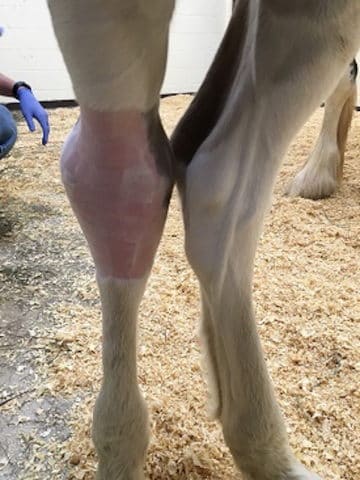
Swollen right hock in septic foal - Classic case: Foal less than 14 days old with lethargy, decreased nursing, +/- obvious septic foci (joint effusion, omphalophlebitis, diarrhea, or pneumonia)
- Dx:
- Blood culture is gold standard but takes 4-7 days
- Increased or decreased neutrophils with bands
- Increased lactate
- Check blood IgG to assess for failure of passive transfer (less than 400 mg/dl)
- Ultrasonography/radiography
- Tx: Broad-spectrum antimicrobials, IV fluids & plasma, anti-endotoxin therapies, nutritional support; treat specific infections (e.g., lavage joint for septic joint, anti-diarrheals for diarrhea, nebulization for pneumonia)
- Pearls:
- Good prognosis at referral centers with aggressive treatment
- CHECK ALL FOALS for adequate passive transfer at 12-24 hours of age to help decrease risk of sepsis
- Gram-negative pathogens most common
- Foals’ conditions deteriorate rapidly so any decrease in nursing or lethargy in a young foal is an emergency!


Images courtesy of John Storr (zebras), Cynthia Powell (corneal ulcer), Nora Grenager (sinus, foal, PPID), Patrick Vermuyten (Arabian stallion).
Top Topic Category
Equine