For my own personal use only:
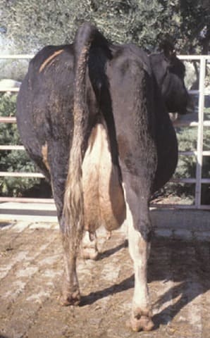
- Bovine lymphosarcoma
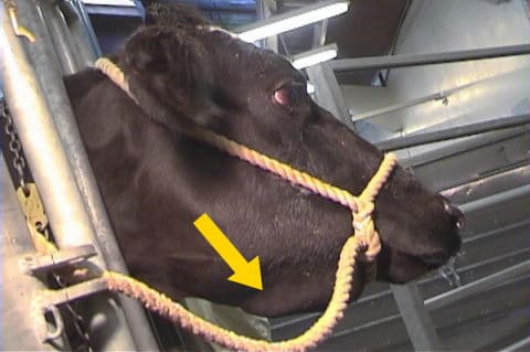
Submandibular lymphadenopathy - Classic case:

- Central nervous system: paraplegia, tetraplegia, paraparesis, tetraparesis; head tilt, facial paralysis, dysphagia
- Gastrointestinal system: free gas bloat, vagal indigestion, palpably enlarged abdominal lymph nodes, melena, thickened rectum
- Lymph nodes: 25% of cases have peripheral lymphadenopathy; can also see exophthalmos, weight loss
- Cardiac: unexpected and sudden collapse, death, congestive heart failure, jugular pulse, distended jugular or mammary veins, arrhythmia, tachycardia, weak pulse, subcutaneous edema
- Dx:
- Etiology: bovine leukemia virus (BLV), an oncogenic retrovirus
- Gold standard: lymph node biopsy and histopathology confirms lymphosarcoma
- BLV infection: Positive antibodies ( ELISA most common) to BLV or PCR or antigen-capture ELISA
- Note: Serologic or virologic positive for BLV does not definitely diagnose lymphosarcoma because prevalence of BLV is high and so few seropositive cases develop lymphosarcoma
- Tx: NO effective or legal treatment
- Control: test and cull positive animals greater than 6 months of age in herds with a low BLV prevalence
- Prevent horizontal spread in herds with high prevalence by changing needles, using rectal sleeves, and good fly prevention
- Pearls:
- Grave prognosis for lymphosarcoma
- Cows w/ lymphosarcoma will not pass slaughter inspection
- Prevalence of BLV in most U.S dairy herds is high, but most are asymptomatic and their prognosis for life is good
- Upwards of 30% of infected animals will have persistent lymphocytosis serving as a reservoir for further spread
- Only approx. 5% of BLV-infected animals get lymphosarcoma
- Classic case:
- Ketosis
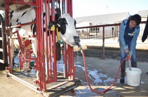
Transfaunation - Classic case:
- 3 types – thin cow up to 45 days post-partum (type I), obese cow peri-partum (type II), or too much silage at any stage of lactation (silage type)
- Dairy cow 1-4 weeks post-partum with mild anorexia, low milk production, malodorous breath
- Pica or paresthesia, aggressive behavior
- Mild proprioceptive deficits with hepatic failure
- Subclinical
- Can be common in dairy herds
- High blood ketone levels without obvious clinical signs
- Decreased milk production and reproductive fertility possible
- Increased risk of metritis, displaced abomasum, early culling from the herd
- Dx:
- Measure betahydroxybutyric acid in blood (best), milk, or urine; w/ clinical ketosis is greater than 14.4 mg/dL in whole blood
- Look for acetoacetic acid in urine with dipstick
- Tx: Depends on type:
- Type I – simple and short term Tx: oral propylene glycol, IV dextrose
- Corticosteroid use is controversial
- Drives glucose into cells but also quells the immune response
- Risky in a post-partum animal
- Type II – difficult and longer-term Tx: transfaunate and pump with liquid nutrition via ororuminal tube, IV dextrose
- Silage type – don’t feed silage with high concentrations of butyric acid to pre- and post-fresh cows!
- Pearls:
- Type I – excellent prognosis; prevent with low-protein diet, maximize energy in early lactation, and monensin
- Type II – poor prognosis; manage dry periods and dry cow nutrition to prevent obese cows, cull
- Economically important because higher culling rates, more retained placentas, decreased pregnancy rates, and decreased milk production in obese cows
- Classic case:
- Bovine viral diarrhea (BVD)
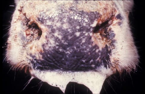
Ptyalism - Classic case:
- Unvaccinated younger cow with acute diarrhea, nasal discharge, ptyalism, ocular discharge, oral ulcers, fever, anorexia, coronitis (not all will occur)
- In-utero infection: early embryonic death, cerebellar hypoplasia, developmental defects, abortion
- Persistent infection (PI): if a cow becomes infected by a NON-cytopathic strain of virus between 40-120 days gestation, or calf is from a PI dam, the calf will be PI and act as a reservoir; if infected by cytopathic strain later in life, cow gets mucosal disease
- Mucosal disease: acute signs and fatal with 2-4 weeks
- Dx:
- Etiology: bovine viral diarrhea virus, a Pestivirus
- Gold standard: virus isolation however PCR on milk, serum, whole blood, tissues or semen is the rapid test of choice
- Antigen-capture ELISA on blood or tissue
- Often requires paired serology to definitively diagnose recent infection vs. exposure or vaccination
- Virus isolation or histopath at necropsy
- Ear notching for PI cattle (PCR or ELISA): most commonly done on calves
- Tx:
- Supportive care for simple adult, unvaccinated cows showing signs of diarrhea, fever and anorexia; prognosis for these animals is good
- None for calves born with developmental defects or animals with mucosal disease
- Prevention: Test and remove PI calves; vaccinate
- Pearls:
- Grave prognosis except for adult with mild, non-mucosal disease
- Worldwide, economically important pestivirus
- Not zoonotic but very contagious
- Classic case:
- Omphalitis, septicemia, joint ill,
meningitis
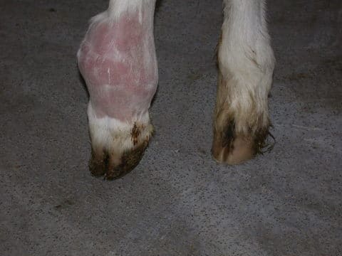
Joint ill - septic metatarsophalangeal joint - Classic case:
- Omphalitis: fever with swollen, painful umbilicus and patent urachus +/- GI pain with secondary peritonitis
- Septicemia: calf less than 2 weeks old with fever, diarrhea, depression, systemic compromise
- Joint ill: lameness with painful, swollen, hot joint
- Meningitis: opisthotonus, hyperesthesia
- Dx:
- Omphalitis: palpate and ultrasound umbilical structures
- Joint ill: ultrasound/radiograph affected joint, aspirate and culture joint fluid
- Meningitis: CSF aspirate shows increased WBC count and protein
- Tx: antimicrobials and…
- Omphalitis: surgical removal for advanced cases
- Joint ill: lavage joint then instill antibiotics; analgesics/NSAIDs
- Meningitis and septicemia: systemic supportive care, NSAIDs, diazepam if seizures
- Prevention: make sure calves get a minimum 500 grams IgG on first feeding and and 4 L colostrum by 4 hours of age; clean calving environment
- Pearls:
- Prognosis variable: good for omphalitis, poor for others
- Measure total protein at 24 hours’ age: adequate colostral transfer if greater than 5.5 g/dL
- Classic case:
- Traumatic reticuloperitonitis
Papple shape - Classic case:
- Acute anorexia and agalactia, unwillingness to move or lie down, arched back, fever, positive grunt test
- +/- Papple shape (pear on right and apple on left) and scant feces if secondary vagal indigestion
- Sloshing fluid sound left thoracic cavity in severe cases with pericarditis
- Rare in pre-ruminating heifers
- Dx:
- Positive withers grunt test: pinch withers while listening for a vocalization with stethoscope
- Look for pain response while balloting left ventral abdomen
- Abdominocentesis: purulent or serosanguineous fluid
- Cranial abdominal ultrasound or radiography
- Tx: Similar outcomes with medical and
surgical,
better prognosis if treated early
- Medical: magnet, laxatives, antimicrobials, analgesics
- Surgical: rumenotomy, antimicrobials, magnet
- Prevention: ONE magnet per cow given at 400-600 pounds weight can prevent
- Pearls:
- 75% survival; bad outcome with secondary vagal indigestion, diffuse peritonitis, and pericardial involvement
- Classic case:


Images courtesy of Dr. Lisle George and The Yorck Project (3000-year old image of a farmer plowing with cattle, Egypt)
Top Topic Category
Ruminants