For my own personal use only:
- Dilated cardiomyopathy (DCM)
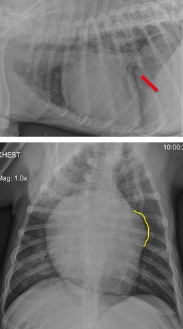
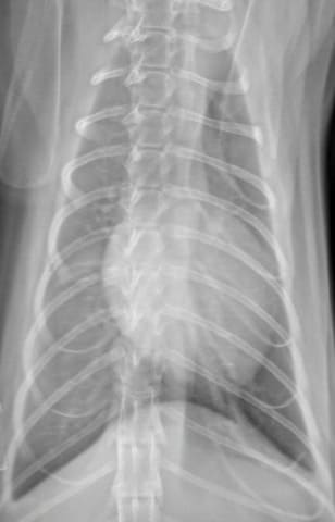
Thoracic radiographs of a boxer with DCM demonstrating left atrial and ventricular enlargement. Top: Mild perihilar infiltrate (red arrow) consistent with some pulmonary edema. Loss of cranial cardiac waist is indicative of right heart enlargement. Bottom: Left auricular bulge (outlined in yellow). 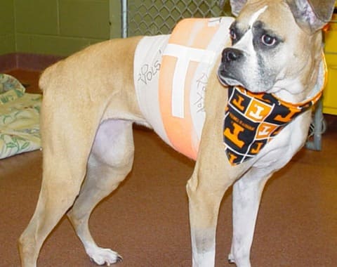
Holter ECG demonstrating runs of ventricular tachycardia with a rate of 274 bpm (black arrow) and 225 bpm (orange arrow) 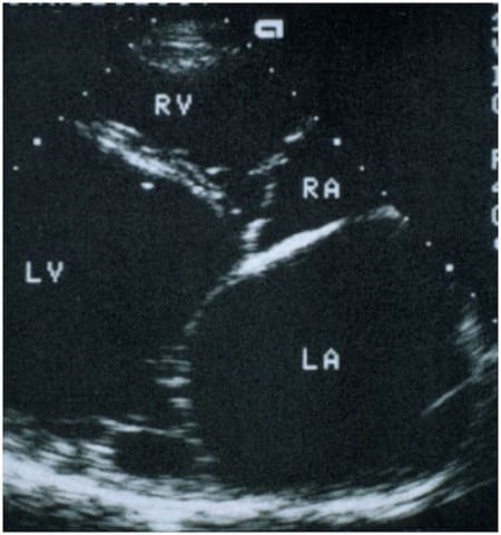
Echocardiogram of a dog with DCM showing marked left atrial (LA) and ventricular dilation (LV), long-axis view - Classic case:
- Large and giant breeds: Doberman pinscher, boxer, great Dane, Irish wolfhound, standard poodle (but also cocker spaniels)
- Middle-aged
- Early signs:
- Heart murmur
- Middle-aged
- Weak peripheral pulses
- Exercise intolerance
- Congestive heart failure (CHF)
- Cough
- Tachypnea and dyspnea
- Tachycardia
- Weakness
- Classic case:
- Dx:
- Radiography:
- Generalized cardiomegaly
- Variable venous dilation
- Pulmonary edema/perihilar infiltrate if decompensated
- Electrocardiography (ECG)/Holter monitor:
- Ventricular ectopic beats are common
- May see significant ventricular arrhythmias early in Dobermans
- Atrial fibrillation in advanced disease
- Holter monitoring to determine:
- Severity of arrhythmia
- If therapy indicated
- Echocardiography:
- Left atrial and ventricular dilation
- Mitral +/- tricuspid valve regurgitation
- +/- Right atrial and ventricular dilation
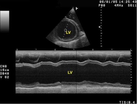
- Evidence of poor contractility:
- Prolonged end-point septal separation (EPSS)
- Decreased fractional shortening (FS)
- Radiography:
- Tx:
- Prior to onset of CHF:
- Angiotensin-converting-enzyme (ACE) inhibitors
- Pimobendan (positive inotrope and vasodilator) if heart dilation present
- Anti-arrhythmics (e.g., sotalol, mexiletine) for ventricular arrhythmias
- After onset of CHF:
- Acute therapy:
- Oxygen
- Stress reduction
- Parenteral furosemide
- Pimobendan
- Chronic therapy:
- Oral furosemide
- ACE inhibitors
- Pimobendan
- Acute therapy:
- Prior to onset of CHF:
- Pearls:
- Prognosis:
- Dogs with occult disease can live several years
- Sudden death due to arrhythmias can occur even before echocardiographic changes of DCM are seen!
- Pimobendan:
- If started prior to onset of CHF will delay development of CHF
- Can extend life during CHF, but prognosis after 1 yr is poor
- Other causes of DCM
- Taurine deficiency-linked in American cocker spaniels, golden retrievers, boxers, and Dalmatians
- Carnitine-responsive in boxers
- Chagas myocarditis caused by the protozoan Trypanosoma cruzi
- Parvovirus due to in utero exposure, rare now
- Doxorubicin-induced caused by cumulative doses greater than 180 mg/m2
- Feline DCM
- Breed predilections: Siamese, Burmese, Abyssinian
- Prognosis is poor because most present in CHF
- Dx and Tx similar to canine DCM
- Taurine-deficiency (commonly seen prior to 1987; now rare because of increased dietary taurine in most cat foods)
- Prognosis:
- Pericardial effusion (PE)
- Classic case:
- Acute onset of weakness/collapse
- Exercise intolerance
- Abdominal distention
- Muffled heart sounds
- Tachycardia
- Weak femoral pulses
- Jugular pulses
- Pulsus paradoxus: Increase in pulse pressure during expiration and decrease during inspiration (normally occurs, but not as easily palpable as during PE)
- Pale mucous membranes
- Dx:
- Etiology: Seen in certain dog breeds depending on underlying
cause:
- Cardiac tumors: German shepherd, golden retriever, boxer, Labrador retriever, bulldog, Boston terrier
- Idiopathic PE: Golden retriever, Labrador retriever
- ECG:
- Tachycardia
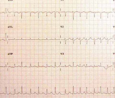
- Electrical alternans: Beat to beat alternating height of R-wave amplitude (due to swinging motion of heart within fluid-filled sac) occurs in 50% of patients
- Low R-wave amplitude
- Radiography:
- Rounded, globoid cardiac silhouette
- Dilated caudal vena cava
- Ascites due to passive congestion
- Echocardiography:
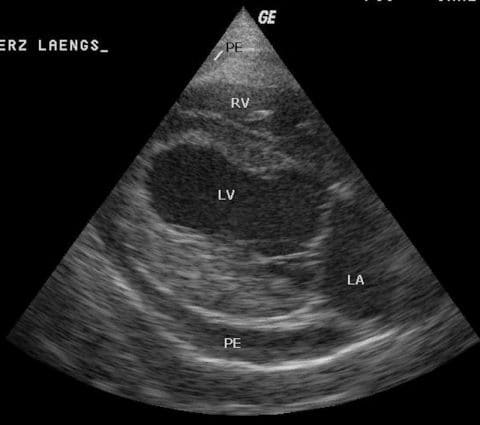
- Fluid-filled sac surrounding heart
- Cardiac tamponade: Flailing right heart due to pressure from PE
- Possibly a causative mass in right atrium, right auricle, or at heart base
- Cytology of effusion:
- Unless due to lymphoma or infection, it is rarely is of diagnostic value
- Effusion PCV of less than 10% appears to improve the diagnostic yield
- Etiology: Seen in certain dog breeds depending on underlying
cause:
- Tx:
- Ultrasound-guided pericardiocentesis:
- Use closed collection system
- Typically on right between 4th and 6th ribs at costochondral junction
- Record ECG during procedure as arrhythmias will be seen if heart is accidentally touched with needle
- Pericardectomy:
- For recurrent effusions to decrease risk of tamponade
- Can be curative for idiopathic PE
- Treatment of neoplasia:
- Right auricular hemangiosarcoma: Possible surgical resection
- Lymphoma, chemodectoma, hemangiosarcoma, and mesothelioma: Chemotherapy
- Ultrasound-guided pericardiocentesis:
- Pearls:
- Prognosis:
- Idiopathic: Good if effusion rarely recurs or if pericardectomy
- Neoplastic:
- Hemangiosarcoma and mesothelioma:
- Poor long-term prognosis
- Recurrent effusions and metastatic disease are most common cause of death
- Chemodectoma:
- Guarded long-term prognosis
- Pericardectomy can prolong survival if PE recurrent
- Hemangiosarcoma and mesothelioma:
- Ultrasonography is diagnostic AND can identify underlying causes such as tumors
- PE in cats is usually a manifestation of heart failure
- Prognosis:
- Classic case:
- Heartworm disease
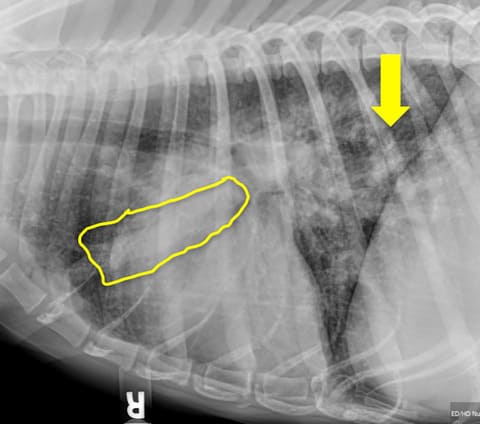
Radiograph of heartworm-positive dog showing a dilated pulmonary artery (yellow tracing) and pulmonary parenchymal disease (yellow arrow) - Classic case:
- Canine:
- Mild:
- No clinical signs
- Cough
- Moderate: Mild signs plus exercise intolerance
- Severe: Moderate signs plus ...
- Dyspnea
- Abdominal distension
- Caval syndrome: Severe signs plus ...
- Acute onset of lethargy or weakness
- Coffee-colored urine due to hemoglobinuria
- Mild:
- Feline:
- Vomiting
- Intermittent cough
- Increased respiratory rate
- Sudden death
- Canine:
- Dx:
- Etiology: Dirofilaria immitis
- Canine:
- Heartworm antigen SNAP test:
- Recommended yearly
- Detects protein secreted by female worm
- Earliest can be detected is 5 mos post-infection (PI)
- Potential causes for false negatives:
- Antigen-antibody complex formation
- Immature females (early infection)
- Light infection
- Male-only infection
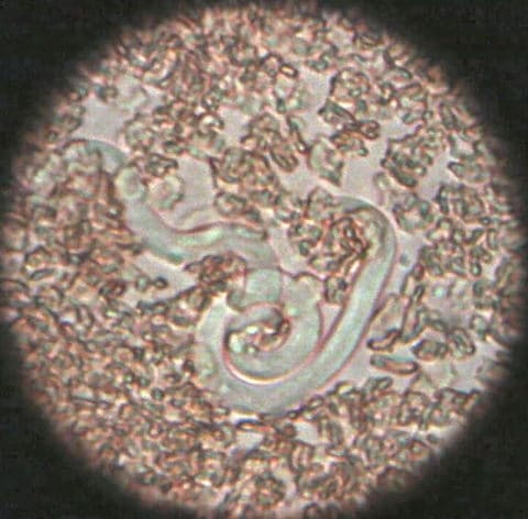
- Microfilaria tests:
- Recommended yearly concurrent with antigen testing
- 7% of infected dogs will be negative on antigen testing and positive on microfilaria testing
- Options:
- Modified Knotts
- Filter test
- Direct smear of anti-coagulated blood
- Thoracic radiography to show degree of
cardiopulmonary disease:
- Enlarged, tortuous, +/- blunt-ending pulmonary arteries
- Right heart enlargement
- Pulmonary parenchymal disease
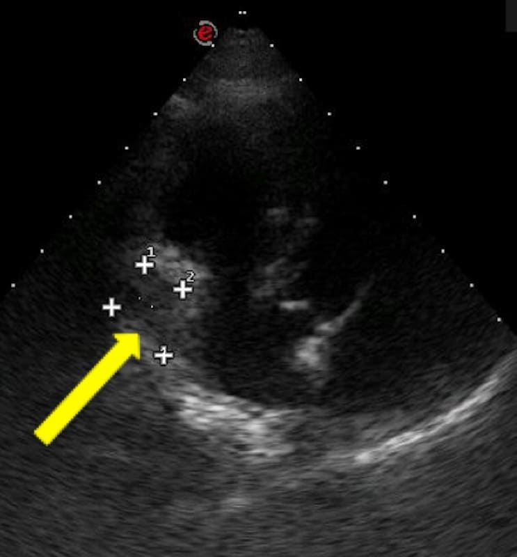
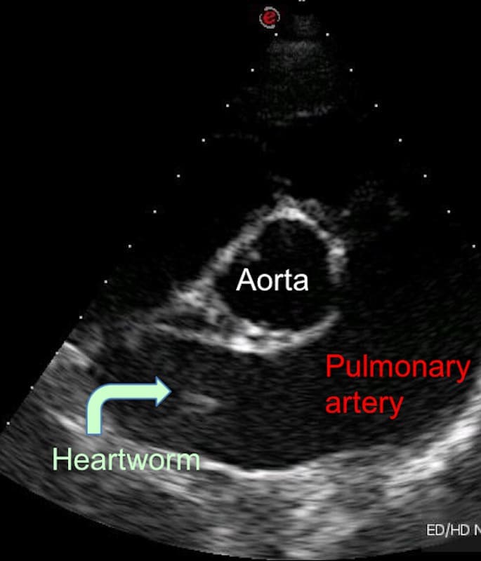
- Ultrasonography: May see worm in pulmonary artery
- Heartworm antigen SNAP test:
- Feline:
- Radiography:
- Only 50% of cats have suggestive radiographic changes as seen in dogs
- May see patterns similar to bronchitis or asthma
- Antigen testing: False negatives if male-only infection, immature infection, or low worm burden
- Antibody testing: Positive only tells you infection has occurred at some time
- Ultrasonography: May see worm in pulmonary artery
- Radiography:
- Tx:
- Canine:
- Doxycycline:
- Daily treatment for 30 d before adulticide therapy
- Reduces Wolbachia : Gram-negative, intracellular bacteria essential for worm survival
- Macrocyclic lactones/heartworm preventatives:
- Start 2 months before adulticide therapy
- Prevent new infections
- Eliminate susceptible larvae and microfilaria (do NOT kill adults)
- Allow immature worms to mature to a stage more susceptible to adulticides
- Pre-treat with diphenhydramine and corticosteroids if microfilaria-positive to prevent anaphylaxis secondary to rapid die-off
- Adulticide therapy:
- Melarsomine dihydrochloride
- 3-dose protocol recommended for all
stages
of heartworm disease:
- IM dose once
- After 1 mo give 2 doses 24 h apart
- Kills 98% of worms
- STRICT exercise restriction starting with first dose of adulticide and continuing for 6-8 wks after last dose
- Corticosteroids:
- Tapering anti-inflammatory dose to control clinical signs of pulmonary thromboembolism
- Start corticosteroids with doxycycline and macrocytic lactone if symptomatic or microfilaria-positive
- Start corticosteroids with first injection of adulticide
- Surgical extraction of adult worms:
- Indicated for dogs with caval syndrome
- Typically requires referral
- Doxycycline:
- Feline:
- Melarsomine NOT recommended
- Prednisolone to help with lung changes
- Canine:
- Pearls:
- Canine:
- Prognosis:
- Good for mild-moderate cases
- Fair to guarded for severe cases
- Poor to grave for caval syndrome
- Prognosis:
- Feline:
- Prognosis is guarded to fair
- Canine:
- Classic case:
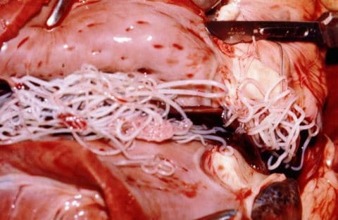
Images courtesy of Laura Cousins DVM, MS, DACVIM (echocardiogram of DCM, radiographs of DCM, Holter ECG of DCM with ventricular tachycardia, radiograph of cat with PE, echocardiogram of PE with mass, echocardiogram of adult heartworm, radiograph of heartworm disease), James Heilman, MD (electrical alternans in a human with PE), Alan R Walker (adult heartworms in heart), Joelmills (microfilaria), and Kalumet (M-mode of DCM and PE echocardiogram) .
Top Topic Category
Canine
