For my own personal use only:
6. Mycoplasma hyorhinis and M. hyosynoviae
- Classic case: both can cause lameness/swollen joints
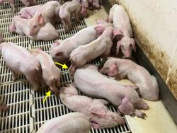
- M. hyorhinis affects nursery pigs (three to 10 weeks of age)
- Unthrifty pigs post-weaning
- Head tilt: Otitis media
- Lameness and swollen joints
- Cough
- M. hyosynoviae affects finishing pigs (10 to 20 weeks of age)
- Acute lameness with or without joint swelling
- Up to 50% mortality
- M. hyorhinis affects nursery pigs (three to 10 weeks of age)
- Etiology
- M. hyorhinis grows better in culture than other types of Mycoplasma, others are too slow-growing
- Dx
- Gross lesions: think thickened tissues
- M. hyorhinis
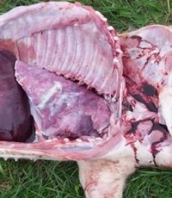
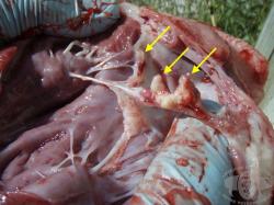
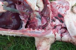
- Fibrinous pleuritis, pericarditis, and sometimes peritonitis
- Thick serosal membranes and fibrinous adhesions
- M. hyosynoviae
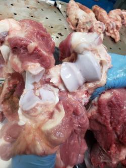
- Thick, edematous synovial membranes and joint structures
- Increased volume of synovial fluid (± brown or cloudy)
- M. hyorhinis
- Microscopic lesions
- M. hyorhinis: Mycoplasma may be visualized on the cilia of the inner ear
- M. hyosynoviae: Perivascular infiltration of lymphocytes, plasma cells, macrophages
- For both: PCR on joint fluid
- M. hyorhinis
- Swabs of serosal surfaces or joints (not lungs)
- Can culture joint fluid (pre-mortem sample)
- M. hyorhinis
- Gross lesions: think thickened tissues
- Tx
- Both: Injectable antimicrobials
- Tylosin
- Lincomycin
- Early Tx for M. hyorhinis is effective but advanced Dz is refractory
- Low mortality rate for M. hyosynoviae
- Both: Injectable antimicrobials
- Pearls
- M. hyorhinis
- Ubiquitous organism in the porcine respiratory tract
- Disease results from invasion and systemic proliferation of the organism
- Clinically similar to Glaesserella and Streptococcus
- M. hyosynoviae is not found in pigs < 4 wks of age, OCD may predispose
- Similar presentation to Erysipelas but will not respond to Tx with penicillin
- M. hyorhinis
7. Glaesserella parasuis (a.k.a. “Glässer disease” and previously called Haemophilus parasusis)
- Classic case
- Ages affected: Nursery pigs (three to 10 weeks of age)
- Sudden death
- Fever
- Cough
- Neurologic signs: Head tilt
- Lameness and swollen joints
- Wasting/unthrifty pigs
- Mortality is high once showing signs if delay or fail to provide individual Tx
- May find suddenly dead pigs in some cases
- Ages affected: Nursery pigs (three to 10 weeks of age)
- Etiology
- Small gram-negative rod with many serovars
- Hard to grow in lab
- Dx
- Gross lesions: Fibrinous polyserositis of the peritoneum, pericardium, and pleura
- Microscopic lesions
- Polyserositis with fibrinopurulent exudate consisting of fibrin, neutrophils, and macrophages on serosal surfaces
- Fibrinopurulent meningitis
- PCR is best since it is difficult to culture
- Try culture of locations where the microbe is not expected
- Serosal surface
- Exudate
- Tx
- Prompt injection of appropriate antimicrobials
- Ceftiofur
- Enrofloxacin
- Tulathromycin
- Vaccination
- Piglets twice
- Sows pre-farrowing
- Prompt injection of appropriate antimicrobials
- Pearls
- Commonly diagnosed cause of poor nursery pig performance
- Prognosis depends on the speed of Tx
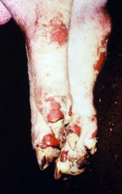
NOTE: Signs of Seneca Valley virus are clinically INDISTINGUISHABLE from multiple reportable diseases in pigs, including foot and mouth Dz (FMD), swine vesicular Dz, and vesicular stomatitis, so quarantine and notify the state vet ASAP for testing and guidance
- Classic case
- Any age animal
- Typical outbreaks in sows (with stress): Lameness
- Cases peak in summer
- Multifocal round erosions or vesicles on distal limb (coronary band), snout/nares, lips/oral mucosa
- Etiology: Picornavirus, genus Senecavirus
- Dx
- Gross lesions as described above
- Microscopic lesions: Lesions seen in the stratified squamous epithelium
- Virus isolation or PCR on serum, oral fluids, vesicles, or vesicle swabs
- Tx
- There are no known treatments or control measures
- Must report vesicles in regions free of foot and mouth disease
- Pearls
- Emerging disease of swine
- Prognosis is usually good but may cause high mortality in neonates
- Classic case
- Ages affected: Farrowing room to nursery (one to 10 weeks of age)
- Cough
- Neurologic signs: head tilt, seizures
- Swollen joints and lameness
- Ages affected: Farrowing room to nursery (one to 10 weeks of age)
- Etiology
- Multiple capsular types
- Facultatively anaerobic, gram-positive, nonmotile coccus (chains)
- Dx
- Gross lesions
- Fibrinous polyserositis
- Vegetative valvular endocarditis (differentiates from Glaeserella)
- Microscopic lesions
- Suppurative bronchopneumonia
- Neutrophilic meningitis or encephalitis
- Fibrinopurulent or suppurative epicarditis
- Interstitial pneumonia secondary to septicemia
- Culture from tissue other than lung, especially protected spaces, e.g.:
- Brain
- Joint
- Pericardial sac
- Gross lesions
- Tx
- Injectable antimicrobials: Ceftiofur, enrofloxacin
- Injectable steroids
- Reported mortality depends on treatment: Can range from 2-20%
- Prevention!
- Use clean tools for tail docking and castration
- Keep farrowing rooms clean
- Maintain good ventilation
- Pearls
- ZOONOTIC: Re-emerging human pathogen: septicemia, meningitis, permanent hearing loss, endocarditis, arthritis
- Common agent in nursery pigs
- May be found in pigs with pneumonia
- More common at times of high humidity
- Prognosis is good with prompt Tx but poor once animals are showing neurologic signs
10. Sarcoptic mange
- Classic case
- Pruritus
- Decreased growth rate
- Etiology
- Burrowing mite: Sarcoptes scabiei
- Entire life cycle is on the skin
- Sows are reservoirs and pass it to piglets
- Demodectic mange is unimportant in swine
- Dx
- Gross lesions
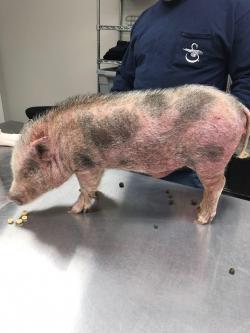
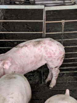
- Erythematous skin
- Progresses to papules on the rump, flank, and abdomen
- Alopecia and abrasions from scratching
- Microscopic lesions
- Papules contain eosinophils, mast cells, and lymphocytes
- Identify the mite on scrapings from inside ear
- Gross lesions
- Tx
- Acaricides
- Injectable: Ivermectin, doramectin
- Topical: Permethrin
- Eliminate from breeding stock
- Acaricides
- Pearls
- Rare in confined herds in the U.S.
- Good prognosis
- DDx may include sunburn
11. Staph. hyicus a.k.a. “greasy pig disease” or “exudative epidermitis”
- Classic case
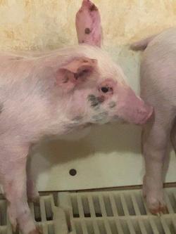
- Starts with focal red areas and clear exudate in groin or on face
- Progresses to coalescing lesions with a thick brown exudate
- Eventually exudate will be thick, black, with a layer of crust over thick, wrinkled skin
- Etiology
- Gram-positive cocci
- Normal skin flora
- Exfoliative toxins linked to virulence
- Dx
- Gross lesions
- As described above plus lymphadenopathy
- Microscopic lesions
- Serocellular crusts of neutrophils and fibrin
- Epidermis is ulcerated and/or hyperplastic
- Dx based on typical appearance of lesions + culture of lesions
- Gross lesions
- Tx
- Early Tx with antimicrobials can be successful, though may be resistant
- None labelled for Staph. hyicus so base choice on susceptibility
- Topical sprays containing chlorhexidine and mineral oil
- Prognosis: Good if disease is mild and treated early
- Poor if other underlying factors are present, e.g.: viruses, poor husbandry, gilt litters (young mothers, poor colostrum)
- Early Tx with antimicrobials can be successful, though may be resistant
- Pearls
- Most common staphylococcal skin disease of pigs
12. Erysipelas rhusiopathiae a.k.a. “diamond skin disease”
- Classic case
- Acute
- Septicemia resulting in lethargy, fever, painful joints, decreased feed intake, classic diamond-shaped skin lesions
- Subacute
- Milder version of acute form
- Chronic
- Follows acute/subacute infections
- Chronic arthritis w/ enlarged hock/stifle/carpus
- Acute
- Etiology
- Gram-positive rod, facultative anaerobe
- Several serotypes
- Dx
- Gross lesions
- Multifocal raised rhomboid/square/diamond-shaped, red to purple skin lesions
- Vegetative valvular endocarditis
- Petechiae on renal cortex
- Microscopic lesions
- Blood vessels in dermis and other tissues: Dilated and congested with bacterial emboli that occlude vessels
- Leads to focal necrosis
- Culture affected tissues with histopathologic lesions
- Gross lesions
- Tx
- Injectable antimicrobials in acutely affected pigs
- Penicillin
- Lincomycin
- Tylosin
- Vaccination
- Sow twice at pre-breeding and at each weaning
- Piglets twice
- Injectable antimicrobials in acutely affected pigs
- Pearls
- Outbreaks in pigs may occur cyclically (every 10 years or so)
- ZOONOTIC
- Good prognosis with prompt Tx
- Tx of chronic cases is difficult and rarely cost-effective so often the best option is to cull
Bonus! - Trichinellosis
- Classic case: No clinical signs in pigs - all about the zoonotic threat
- Etiology
- Several species exist in different regions
- T. spiralis in North America and Europe
- Dx
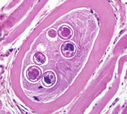
- Histopath of muscle tissue: ID cysts (in diaphragm)
- ELISA
- Tx: None for swine
- Focus on transmission
- Cook pork to 145°F(63°C)
- Regulations for garbage feeding in swine
- Focus on transmission
- Pearls
- ZOONOTIC: Via ingestion of infected muscle tissue
- More common from other sources than pork like consumption of bear meat
All images courtesy of Meghann Pierdon, VMD, DACAW, except where noted - Trichinella histopath (U.S. Centers for Dz Control) and the vesicular lesions on the feet (USDA).
Top Topic Category
Porcine
