For my own personal use only:
1. Porcine reproductive and respiratory syndrome (PRRS)
- Classic case
- Can be subclinical: i.e., infected pigs w/ NO signs
- Breeding herd: Varies from mild to severe
- Abortions
- Early farrowing
- Anorexia
- Up to 100% neonatal mortality
- Growing pigs: Transient disease w/ up to 20% mortality
- Cough, fever
- Secondary infections
- Streptococcus suis
- Glaeserella parasuis
- Mycoplasma spp.
- Etiology
- RNA Arterivirus: Different strains
- Invades pulmonary alveolar macrophages
- Dx
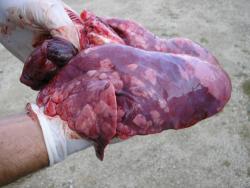
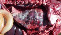
- Necropsy: Lungs fail to collapse w/ multifocal consolidation, enlarged lymph nodes
- Histopath: Necrotizing interstitial pneumonia, lymphoid hyperplasia, and focal follicular necrosis
- Oral fluids or serum on sows and newly weaned piglets
- ELISA
- PCR
- PCR on lungs
- Tx: None
- Supportive: Prevent secondary infections
- Very common: If low level and not causing significant economic losses, may do nothing but monitor
- Pearls
- Most economically costly disease in pig production
- Highly infectious and contagious
- Biosecurity is key!
- Must test new animals before entry
2. Influenza A virus (IAV)
- Classic case: Affects all ages!
- Acute: Transient disease, worse in younger pigs, better in vaccinated pigs
- Sudden onset of fever and cough
- 100% morbidity
- Nasal discharge
- Endemic IAV in sow farms and nursery pigs
- Poor reproductive performance
- Piglets coughing in farrowing crates
- Cough and poor performance in nursery
- Acute: Transient disease, worse in younger pigs, better in vaccinated pigs
- Etiology
- Influenza A viruses
- Major surface antigen types in swine: H1N1, H3N2
- Dx
- Gross lesions:
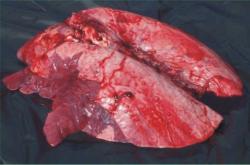
- Sharply demarcated multifocal areas of consolidation
- Microscopic lesions:
- Degeneration and necrosis of the epithelium in the bronchi and bronchioli
- Hyperemia and dilatation of the capillaries
- Infiltration of alveolar septae with lymphocytes etc.
- Oral fluids: PCR (or, less commonly, virus isolation)
- Nasal swabs on febrile pigs for PCR
- PCR on lung tissue
- Gross lesions:
- Tx
- Outbreak
- Supportive care such as aspirin, NSAIDs like flunixin meglumine
- Need to get fevers down so pigs eat and drink
- Treat secondary bacterial infections with antimicrobials
- Prevent via vaccination: sows and piglets
- Homologous protection is best
- Outbreak
- Pearls
- Zoonotic: Humans and other animals can get IAV from pigs so we DON'T call it "swine flu" anymore
- Conversely, pigs are good incubators of different flu viruses they get from birds and humans
- In 2009, an H1N1 IAV strain of swine origin spread globally, infecting people, poultry, dogs, cats, and other animals
- One of the most common respiratory pathogens of growing pigs
- Zoonotic: Humans and other animals can get IAV from pigs so we DON'T call it "swine flu" anymore
- Classic case
- Acute death
- Affects all ages from sows and neonates to finishing pigs
- May be accompanied by cough, lethargy, epistaxis, and sometimes discoloration of ears
- Respiratory disease in finishing pigs
- Dyspnea
- Acute death
- Acute death
- Etiology: Ubiquitous small gram-negative rod
- Dx: Culture visible lesions
- Gross lesions
- Petechial to ecchymotic hemorrhages in multiple organs
- Serous to serofibrinous exudates in the thoracic and abdominal cavities
- Pleuritis, pericarditis, arthritis, and miliary abscesses in a variety of organs
- Microscopic lesions
- Foci of necrosis in multiple organs associated with bacterial thromboemboli
- Gross lesions
- Tx
- Antimicrobials
- Spreads nose to nose so inject antimicrobials into those pigs that have nose-to-nose contact with the pigs that died
- Follow up with antimicrobials via the water if needed
- Good prognosis with Tx
- Pearls
- Can be primary pathogen, but also associated with viral diseases
- Sporadic and difficult to prevent
4. Porcine circovirus diseases (PCV2)
- Classic case
- Ages affected: Pigs early in grower (>10 weeks of age) have two syndromes
- Post-weaning wasting multisystemic wasting syndrome (PMWS)
- Diarrhea
- Porcine dermatitis and nephropathy syndrome (PDNS)
- Pale to icteric skin with coalescing raised red to purple lesions covering the rump
- Post-weaning wasting multisystemic wasting syndrome (PMWS)
- Ages affected: Gilts in sow herd
- Increased mummified fetuses
- Late term abortions
- Ages affected: Pigs early in grower (>10 weeks of age) have two syndromes
- Etiology: Small, nonenveloped DNA virus
- Dx
- Gross lesions:
- Enlarged lymph nodes
- Lungs do not collapse, with interlobular edema
- Kidneys are enlarged and pale and subcapsular surface may have spotted white foci
- Microscopic lesions:
- Lymphocytic histiocytic infiltration of lymphoid tissues
- Sloughing of lung epithelium with fibroplasia
- Oral fluids: PCR
- IHC and PCR (low cycle time [CT]) on lung tissue and lymph nodes
- Gross lesions:
AND
- Histopathologic lesions of lymphocytic histiocytic infiltration of lymphoid tissue
- Tx
- No Tx
- Prevent via vaccination
- Prognosis poor in unvaccinated pigs
- Pearls
- UNcommon now due to vaccines, but can occur in unvaccinated or under-vaccinated pigs
- Ubiquitous in pigs world-wide
- There are other types of PCV that are not pathogenic, or their pathogenicity is unknown
5. Mycoplasma hyopneumoniae a.k.a. enzootic pneumonia
- Classic case
- Ages affected: Late finishing (>15 weeks of age)
- Deep, barking and/or non-productive cough
- Pigs stop growing
- May occur in sow farms if positive animals are introduced to a negative farm
- Ages affected: Late finishing (>15 weeks of age)
- Etiology: slow-growing bacterium
- Dx
- Gross lesions:
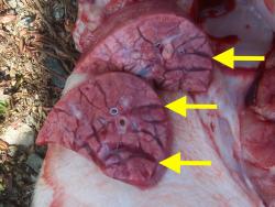
- Cranioventral consolidation (apical, cardiac, and accessory lobes)
- Microscopic lesions:
- Lymphocytes in perivascular, peribronchial, and peribronchiolar tissues
- Cuffing and lymphoid hyperplasia around the airways
- Not a good candidate for culture
- PCR: Lung, oral fluids, tonsil scrapings
- ELISA in negative herds
- Gross lesions:
- Tx
- Prevent using vaccination
- In piglets (twice)
- In replacement gilts depending on status of sow herd they are entering
- Antimicrobials during outbreaks in late finishing pigs
- Hard to eliminate cough in pigs
- Prevent using vaccination
- Pearls
- May be seen in conjunction with PRRS or other pathogens
- Many farms have eliminated the pathogen
- Poor growth may persist after infection
Bonus! - Atrophic rhinitis
- Classic case: Ages affected: three- to six-week-old pigs
- Sneezing
- Nasal discharge
- Tear staining
- Decreased growth rate
- Etiology: Cause is combination of Bordetella bronchiseptica ± toxigenic Pasteurella multocida + management factors (e.g., poor air quality)
- Dx
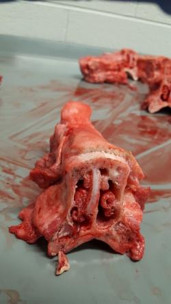
- Gross lesions
- Mild to moderate turbinate atrophy
- ± Deviation of the nasal septum
- Microscopic lesions
- Disruption of normal bone formation in the turbinates
- Gross lesions
- Mild to moderate turbinate atrophy
- ± Deviation of the nasal septum
- Microscopic lesions
- Disruption of normal bone formation in the turbinates
- Gross lesions
- TX
- Vaccination
- Sow twice @ pre-farrowing
- Piglets twice
- Antimicrobials to the sow at parturition
- Vaccination
- Pearls
- No longer a major health concern in the US - RARE
- Still found at slaughter surveillance
- Usually mild Dz and turbinate damage may be reversible
All images courtesy of Meghann Pierdon, VMD, DACAW.
Top Topic Category
Porcine
