For my own personal use only:
- Foot-and-mouth disease
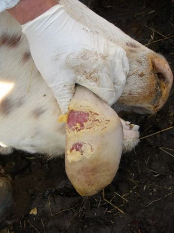
Eroded FMD ulcers on bovine tongue 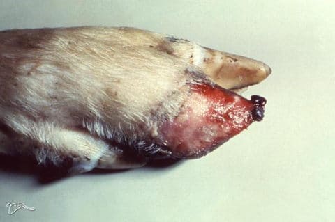
Detachment of horny tissue of a pig's trotter following rupture of FMD vesicles - Classic case: Cattle, sheep, and pigs (NOT
horses)
- Drooling: Viscous, sticky saliva; lip smacking; bruxism
- High fever
- Lameness: Foot stomping, shifting feet
- Painful vesicles or erosions on tongue, muzzle, gums, teats, between claws, coronary bands
- Poor milk production, agalactia
- Abortions, neonatal disease/death
- Spreads quickly!
- Dx: Situation determines which test to use (i.e.,
surveillance, carriers, outbreak)
- Etiology: an Aphthovirus of the Picornavirdae family, 7 serotypes
- ELISA, PCR, virus isolation, electron microscopy (EM),
complement fixation (CF):
- Samples: Vesicular fluid, epithelium, exudates, pharyngeal/esophageal fluid, milk, semen, blood
- Approved labs perform initial testing; special reference labs do confirmatory testing
- Tx: Preventative measures
- Euthanize all positive and in-contact animals; burn or bury carcasses
- Maintain strict movement/entry requirements
- Quarantine +/- vaccination (killed vaccine provides 4-6 mo immunity)
- Thorough disinfection of premises, equipment, etc.
- Use disinfectant with pH less than 6 or greater than 9
- Pearls:
- One of the MOST contagious animal diseases known
- Reportable worldwide
- SEVERE economic impact and production losses
- Export/travel bans on animals and products within and between nations
- Prognosis:
- Good for infected individuals
- Poor for overall herd health and economic outcome
- Guarded in neonates and nursing animals
1½. Vesicular stomatitis (VS, "evil twin" to FMD)
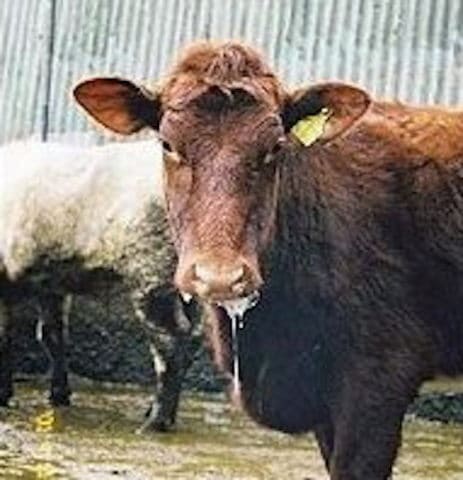
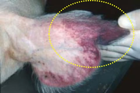
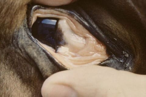
Hypersalivating cow secondary to VS 
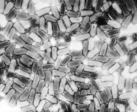
Transmission electron microscopic (TEM) image of numerous VS virus virions 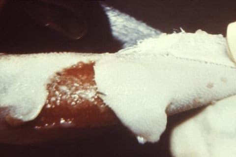
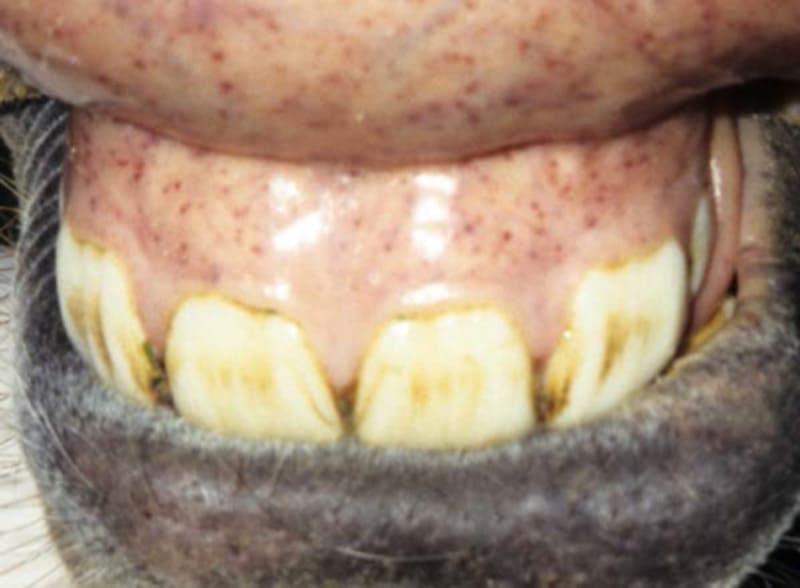
Epithelial detachment of the tongue of an unknown farm animal with VS - Classic case: CATTLE and HORSES, occasionally
swine, camelids, rare in sheep/goats
- Warm humid areas in western hemisphere (Mexico, Central America, parts of South America, Southwest USA)
- Adult animals greater than 1 yr
- Fever
- Salivation, difficulty eating
- Lameness
- Secondary infection (mastitis)
- Vesicles, erosions, ulcers on mouth, lips, teats, udder, coronary bands, sheath, belly
- Hyperemic (skin) or raised blanched areas (oral)
- Morbidity variable, mortality very low
- Dx:
- Etiology: Family Rhabdoviridae, genus Vesiculovirus
- Two serotypes: New Jersey (NJ) and Indiana (IND)
- Transmission by insects (sand/black flies, mosquitos) or direct contact with saliva, epithelium, exudates, or fomites
- Identify viral antigen or antibody (ELISA most commmonly used)
- Etiology: Family Rhabdoviridae, genus Vesiculovirus
- Tx:
- Symptomatic care: Soft feed, bedding, +/- analgesics
- Antibiotics
- Prevention:
- QUARANTINE farm
- Isolate affected animals
- Sanitation/disinfection
- Insect control/exposure
- Vaccines available
- Pearls:
- Zoonotic
- Cannot be distinguished from FMD, swine vesicular disease (SVD), or vesicular exanthema of swine (VE) by clinical signs alone
- Although mortality rare, there is significant economic loss
- The big 8 rule outs of vesicular diseases:
- Bluetongue
- Bovine papular stomatitis (BPS)
- Bovine viral diarrhea (BVD)
- Bovine papular stomatitis (BPS)
- FMD
- Infectious bovine rhinotracheitis (IBR)
- Malingnant catarrhal fever (MCF)
- Rinderpest
- VS
- Classical swine fever (CSF, hog cholera)
Hypermetria or "goose-stepping" in a pig with CSF 
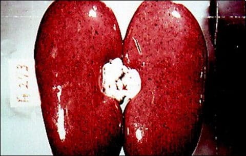
Pinpoint hemorrhages on kidneys are characteristic of CSF 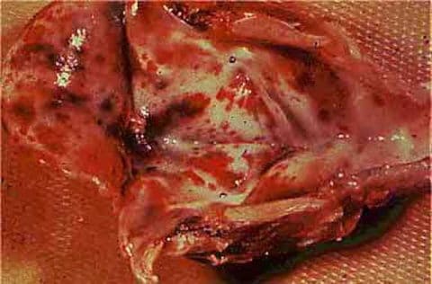
Hemorrhages on larynx of pig with CSF
Image courtesy of The Pig Site- Classic case: PIGS only
- Severity varies with:
- Age: Young animals most severe with high mortality
- Immune status of herd
- Strain
- Acute form (~100% morbidity/mortality):
- High fever >105℉ (>41℃)
- Anorexia
- Constipation followed by watery diarrhea
- Cyanosis, erythema, skin hemorrhages
- Staggering, incoordination, posterior paresis, seizures
- Death within 1-3 wks
- Some cases ASYMPTOMATIC, inapparent carriers
- Severity varies with:
- Dx:
- Etiology: RNA Pestivirus of family Flaviviridae
- Suspect if:
- Septicemia, high fever, incoordination, diarrhea, deaths
- History of feeding garbage, new/returning animals to herd
- NO response to treatment
- Cases on nearby farm
- Necropsy:
- Widespread hemorrhages
- "Turkey-egg" kidneys with pinpoint hemorrhages
- Necrotic foci on intestinal mucosa, larynx, epiglottis
- RT-PCR: Commonly used in CSF surveillance
- Tx: REPORTABLE, DO NOT TREAT CSF positive pigs
- Notify Federal and State veterinarians
- Quarantine farm until definitive diagnosis determined
- Isolate CSF-suspect animals
- Prevention:
- NEVER feed pigs undercooked garbage (swill) or pork products
- Quarantine newly purchased, returning animals for a minimum of 30 d
- Pearls:
- Main sources of infection: Carrier pigs, feeding garbage
- Clinically indistinguishable from African swine fever
2½. African swine fever (ASF, "evil twin" to CSF)
Ear tip hyperemia is a common sign of ASF 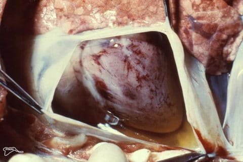
Cut pericarial sac reveals hemorrhages on the myocardium and excess fluid in a case of ASF 
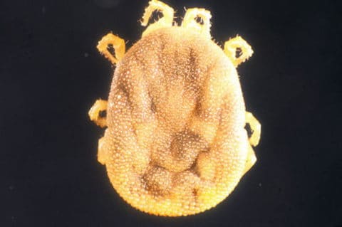
An important vector of ASF, soft, eyeless ticks (Ornithodoros moubata, tampan) inhabit wart hog burrows - Classic case: Acute form (most common)
- Ear tip hyperemia
- Scleral hemorrhage
- Skin cyanosis
- Hemorrhagic intestines
- High fever >105°F (>41°C)
- ~ 100% mortality
- Wart hogs have NO clinical signs (natural host)
- Close contact between domestic pigs and wart hogs (due to infected ticks)
- Dx:
- Etiology: DNA virus, genus Asfivirus. The only member of the family Asfarviridae (African swine fever-like viruses)
- Field diagnosis:
- History and clinical signs
- If suspected, REPORT IMMEDIATELY
- Samples sent ONLY to authorized state diagnostic lab via secure shipping
- Samples: Tonsil (best), kidney, spleen, lymph nodes, whole EDTA blood
- Necropsy: THINK HEMORRHAGIC
- PCR: Tonsil scraping can detect before onset of clinical signs
- Tx: NO treatment
- Quarantine farm
- Slaughter all, burn or bury carcasses
- Prevention:
- Strict biosecurity and sanitation protocols
- Importation restrictions on pigs and pork products
- NEVER feed pigs undercooked garbage (swill) or pork products
- Pearls:
- Vector: Soft ticks that inhabit wart hog burrows
- Devastating economic consequences
- Bovine spongiform encephalopathy (BSE)

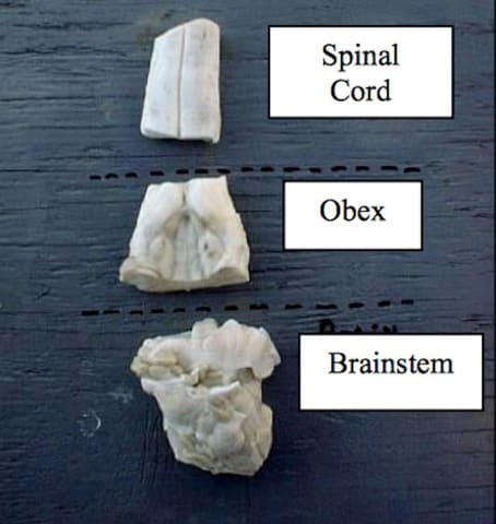
Dorsal view of sections showing the rostral brainstem, obex (best tissue sample for BSE diagnosis), and spinal cord from a cow 
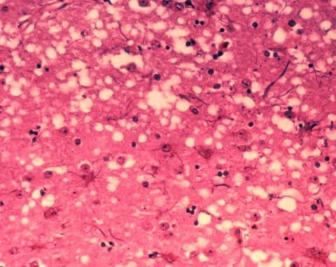
Vacuolar or "spongy" appearance of gray matter in a case of BSE - Classic case:
- Adult animal (> 2 yr)
- Insidious onset abnormal behavior:
- Aggression
- Apprehension
- Ataxia
- Tremors
- Low head carriage
- Weight loss
- Reduced milk yield
- Dx:
- Etiology: BSE is a transmissible spongiform encephalopathy (TSE) caused by a misfolded version of normal cell prion proteins (PrP)
- Screening: ELISA
- If ELISA inconclusive, send to a National Veterinary
Services
Laboratories (NVSL)-approved lab for confirmation:
- Immunohistochemistry (IHC) of obex (in brainstem) +/- electron microscopy
- Western blot used for autolyzed or degraded samples
- List of labs approved by National Animal Health Laboratory Network (NAHLN)
- Tx: None: Euthanasia
- Prevention:
- Do NOT feed animal tissues/products to cattle
- Incineration of carcass is best method to destroy prions
- Test all "downer" cows for BSE
- Take GREAT care in handling tissues
- Prevention:
- Pearls:
- Prognosis: Always fatal
- UNLIKE scrapie (below), pruritus is not a feature of BSE
- Misfolded prion proteins accumulate in cells and cause dysfunction
- BSE is spread by ingestion; no genetic susceptibility required for infection
- BSE is ZOONOTIC: Linked to variant form of Creutzfeldt-Jakob disease (vCJD) in humans
- Scrapie
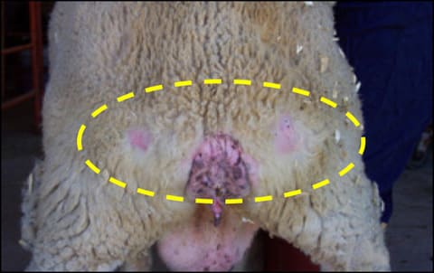
Wool loss over hindquarters from rubbing due to pruritus from scrapie 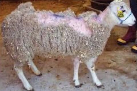
Scrapie in Rambouillet-Cheviot ewe - Classic case:
- Adult sheep (> 2 yr):
- Black-faced breeds in US (96% of cases)
- Many breeds elsewhere
- Seen in goats rarely
- Pruritus is classic; seen in 70% of cases:
- Sheep rub skin constantly
- Wool is scraped off
- Hypersensitivity
- Progressive neurologic signs:
- Head tremors
- Ataxia, bunny-hopping, prancing
- Nibbling at legs and air, lip-smacking
- Behavior changes: Separate from flock, hyperexcitable
- Weight loss with normal appetite
- Death within weeks to months once clinical signs present
- Adult sheep (> 2 yr):
- Dx: Must detect prions proteins in tissue
- Etiology: Mis-folded versions of normal cellular prion proteins (PrPSC)
- Histopathology: Vacuoles, plaques
- IHC is gold standard:
- Brain tissue, most often the obex
- Cerebellum for 'atypical' scrapie
- Western blot when tissues are autolyzed
- ELISA for screening: Brain, lymphatic tissues
- Biopsy lymphoid tissue inside 3rd eyelid for IHC
- Biopsy of tonsils - used in Europe - for IHC or ELISA
- Tx:
- None: Euthanasia
- Prevention: Take GREAT care in handling and transporting
tissues:
- Breed only genetically resistant sheep
- Do not feed ruminant proteins to ruminants
- Maintain closed herds
- Euthanize positive sheep
- FollowEradicate Scrapie guidelines and US Mandatory Scrapie Eradication Program
- Carcasses: Incineration or alkaline digestion
- Pearls:
- Incubation is 2-5 yr
- Prions are normal cellular proteins: No immune response
- Check out these videos on scrapie:
4½. Chronic wasting disease (CWD)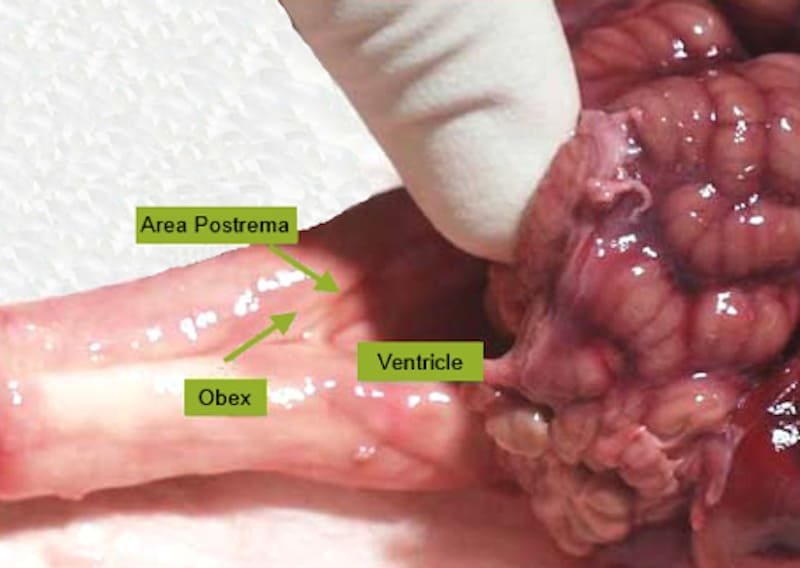
Dorsal view of the obex of the brainstem is seen by lifting cerebellum. The obex is the CNS area most often positive on histopathology testing for CWD 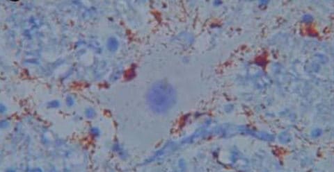
Perineuronal and extracellular deposits of abnormal PrP in CWD - Classic case: White-tailed deer, mule deer, el
- Aspiration pneumonia due to esophageal dysfunction
- Weight loss
- Neurologic signs
- Behavior changes
- Adults older than 16 mos
- Dx: Detect prion protein
- Etiology: Mis-folded versions of normal cellular prion proteins (PrPCWD)
- ELISA for screening
- Confirm with IHC or western blot:
- White-tailed deer and mule deer:
- Brainstem (obex) and lymphoid tissues
- Always submit retropharyngeal lymph node
- Elk: Brainstem (obex) and lymphoid tissues
- White-tailed deer and mule deer:
- Tx: None
- Prevention:
- Quarantine
- Test and cull
- Disposal: Incineration, approved disinfectants
- Prevention:
- Pearls:
- Recently found in Canadian moose
- Prions seen in lymph tissue before CNS in most deer
- Prions very resistant to destruction
- Hunters beware: Cooking DOES NOT destroy PrP
- Horizontal transmission: Direct contact between animals or environment
- Equine infectious anemia (EIA)
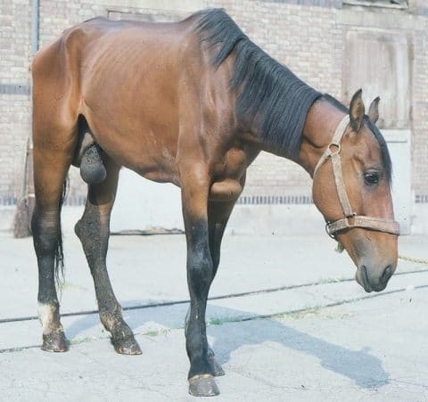
Weight loss and edema (ventral and distal limb) in a horse with EIA 
Pale conjunctiva in a horse with EIA 
Petechial hemorrhages in a horse with EIA - Classic case: Three types
- Inapparent: NO signs (most common form)
- Acute:
- Fever
- Lethargy
- Thrombocytopenia
- Chronic:
- Recurrent episodes of fever
- Anemia
- Weight loss
- Ventral edema
- Petechiation
- Dx:
- Etiology: Lentivirus (Retroviridae family)
- Coggins test: Agar gel immunodiffusion (AGID)
- ELISA
- Testing must be performed at USDA-approved lab and submitted by licensed AND federally accredited veterinarian
- Tx:
- No treatment
- Prevention:
- Seropositive horses must be in lifelong quarantine at least 200 yards from other horses, or euthanized
- All horses moved interstate or sold within a state must have been tested negative for EIA within the last 12 months
- Pearls:
- Lentivirus is related to HIV
- Persistent lifelong infection
- Transmission by biting insects or blood transfer
- Not a common disease in US:
- Historically more common in gulf states (swampy)
- Prognosis: Grave for normal use
- Classic case: Cattle, sheep, and pigs (NOT
horses)


Images courtesy of Craig Packer/USDA (thermography of FMD cow - top image), USDA (ASF ear hyperemia, obex under cerebellum), Izvora (FMD tongue), Joelmills (CSF kidneys), FAO (ASF tick), CDC Public Health Image Library (PHIL) (FMD pig, pig vesicular stomatitis images, ASF heart, goose-stepping pig), USDA APHIS (obex, BSE histopath, scrapie in ewe), Official Nebraska Government Website (cow with VS), Joel C. Watts (CWD histopath), Darreenvt (EIA horse with edema, EIA horse with pale conjunctiva), Dr. Erwin Pearson (petechiae), The Photographer (Charolais - bottom image), and Rachel Keegan (FMD sign) .
Top Topic Category
Cross-Species