For my own personal use only:
- Koi herpes virus (KHV)
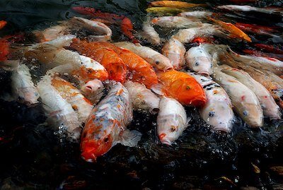
The most popular ornamental species of fish, koi are a variant of common carp 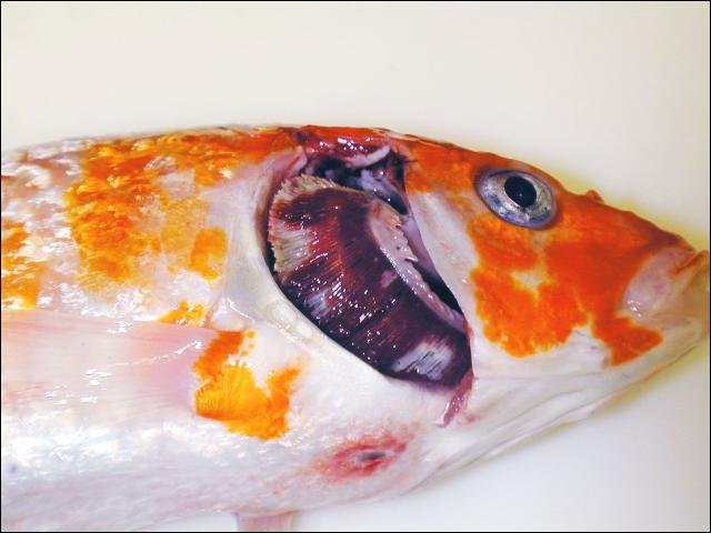
Mottled, necrotic gills, a common finding in koi herpesvirus disease; sunken eyes and a notch in the “nose” may also be seen - Classic case:
- Koi or common carp
- Mottled red/white gills, +/- hemorrhaging, +/- sunken eyes, +/- notched nose
- Surface-swimming, lethargy, respiratory distress
- High morbidity and mortality
- Dx:
- Etiology: caused by Cyprinid herpesvirus-3 (CyHV-3)
- Dx: PCR (ideal for clinically diseased fish) or virus isolation on either fresh dead fish or various tissues (including gill) from very sick fish
- Dx: negative blood samples (for antibodies, i.e., ELISA) do NOT rule out survivor/carrier status and may also be negative for early stage disease
- Tx:
- Depopulation is recommended, but not mandatory: survivors become carriers, perpetuating disease spread
- Some infected koi may be detected with a 30-day quarantine at 75ºF (24ºC) for new arrivals, but carriers may not show clinical signs within this window
- Do not show koi at events that use common/shared containers to display fish
- Pearls:
- KHV is reportable in the U.S. and Canada to their respective federal authorities and to the World Organization for Animal Health (OIE)
- KHV is considered endemic and is common in the U.S. and many other parts of the world
- Goldfish (and a few other species) can be asymptomatic carriers
- Highly contagious
- Classic case:
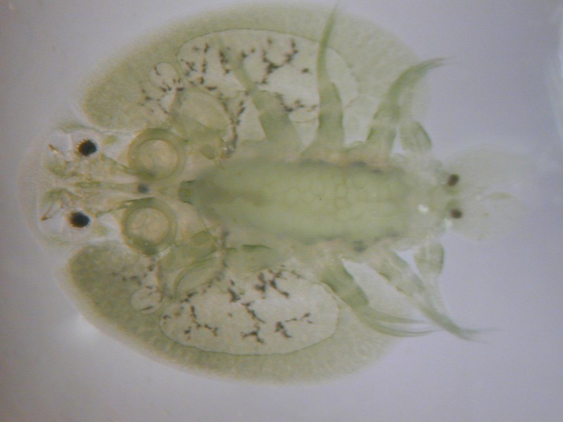
- Fish louse, fish lice, sea lice

Stereomicroscopic shot of Argulus (fish louse) - Classic case:
- Note: the terms fish louse, fish lice, and sea lice refer to a number of different species of freshwater and marine crustacean parasites of fish
- In many cases, adults can be seen grossly and attached, causing skin/gill/fin damage
- Large numbers can cause death
- Dx:
- Sea lice: two of the most common groups include Lepeophtheirus salmonis and Caligus spp.
- Both are copepod crustacean parasites
- Freshwater: Argulus spp. (branchiuran crustaceans) are often seen on freshwater fish (wild game fish, bait fish, goldfish, and koi) in ponds or aquariums
- Dx: use direct visualization but can verify with microscopic examination of the organism
- Sea lice: two of the most common groups include Lepeophtheirus salmonis and Caligus spp.
- Tx:
- Treatment options are limited and will vary depending upon species and situation
- Must consider legalities, human health, and environmental issues in each situation
- Drugs used:chitin/growth inhibitors (affecting juvenile stages), organophosphates; and emamectin, among others
- Secondary bacterial infections may require antibiotics
- Treatment options are limited and will vary depending upon species and situation
- Pearls:
- Lepeophtheirus salmonis is a significant problem in ocean-farmed salmonids and Caligus spp. can infect numerous marine species; the females of both groups hold egg sacs on their bodies
- Argulus species can infect many different freshwater species and lay eggs in the environment
- Argulus has been shown to be a vector for bacteria and viruses
- Classic case:
- Carp/koi pox virus
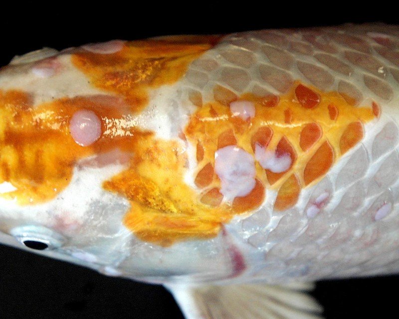
Typical carp/koi pox lesions- smooth, raised, and with a “candle wax drippings” appearance (Photo credit: Andrew Goodwin, USFWS) - Classic case: seasonal
- Benign, non-necrotizing epidermal hyperplasia; lesions are white to cream, smooth or raised; often look like “candle wax drippings” on the fish
- Severe cases can have papillomatous growths +/- secondary bacterial infection
- Dx:
- Etiology: caused by Cyprinid herpesvirus-1 (CyHV-1; related to virus that causes koi herpes virus, CyHV-3)
- History and visual assessment; biopsy/histopathology
- Tx:
- Usually self-limiting, of minimal clinical importance in adults
- May be fatal in fry
- In koi, can cause aesthetic issues and decrease the value of the fish; may need to cull
- Most lesions regress at water temps above 68° F (20°C)
- Usually self-limiting, of minimal clinical importance in adults
- Pearls:
- Carp pox is one of the oldest known diseases of fish
- Prevent with good husbandry and biosecurity
- If feasible, increasing temperatures (e.g., in a hospital tank or system) will help lesions regress
- Classic case: seasonal
- Furunculosis or "ulcer disease"
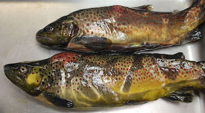
Brown trout with furunculosis - Classic case: in salmonids
- Acute: appears like a bacterial hemorrhagic septicemia; may have hemorrhages in gills, fins, tail, muscles, and internal organs
- Chronic: focal swelling, hemorrhage, and tissue necrosis in muscles
- In some instances, progresses to “furuncles” - deep abscesses that ooze out the skin
- Liquefactive necrosis in spleen, liver, kidney
- Atypical strain: in non-salmonids (but may also infect salmonids)
- Dx:
- Etiology: Aeromonas salmonicida is a gram-negative, nonmotile rod (distinct from the “motile” Aeromonas species that are more common in and/on fish and systems)
- “Sub-species” or “strain” variants will vary and determine species susceptibility and/or clinical signs (i.e., typical vs. atypical presentations)
- Dx: isolate a pure culture from tissue or blood samples or use PCR, especially for atypical A. salmonicida strains that are primarily epidermal (rapid overgrowth of secondary bacterial species impairs culture results)
- Etiology: Aeromonas salmonicida is a gram-negative, nonmotile rod (distinct from the “motile” Aeromonas species that are more common in and/on fish and systems)
- Tx:
- Treat with antibiotics based on culture/sensitivity (C/S) and legal access (e.g., food and game fish species)
- Ulcer disease presentation and Dx by PCR may preclude C/S so use best judgement forTx
- Pearls:
- Important condition in salmonids, goldfish, and koi, along with other freshwater and marine species
- A. salmonicida vaccines available and used for salmonids; mixed results with use of “ulcer disease” atypical strain vaccines (e.g., for koi)
- Classic case: in salmonids
- Mycobacterium
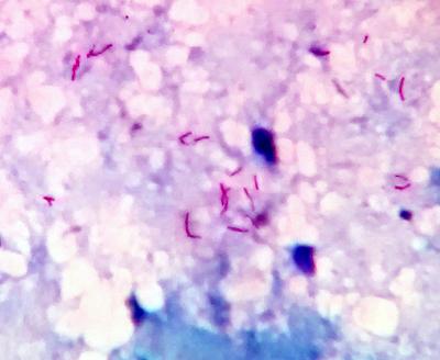
Acid-fast mycobacteria seen under the microscope - Classic case:
- Low-level mortalities, over weeks or months within a population
- In aquaculture or aquarium, may be associated with older or more immunosuppressed (stressed) fish, poor water quality, high organic load, poor husbandry, cannibalism
- “Poor doer” signs - typically over a prolonged period (weeks to months) may share similarities with systemic bacterial or viral infections:
- Coelomic swelling, “pine-coning” (lepidorthosis), exophthalmia
- Skin ulcerations, hemorrhages
- Weight loss, death
- Dx:
- Etiology: Mycobacterium spp. (common - M. marinum, M. fortuitum, and M. chelonae; but other species of mycobacteria can also cause this disease)
- Dx at necropsy: see internal (and/or external) nodules, granulomas, gray-white, necrotic foci in multiple organs; usually see first in spleen and kidney
- In some cases, may see caseous- or abscess-like lesions, hyperpigmentation, and/or no evidence of granulomas or nodules internally (i.e., histiocytic vs. granulomatous (“nodular”) inflammation)
- Dx: acid-fast rods on cytology/histopath
- PCR, culture followed by ID
- Tx:
- None - antibiotics are ineffective
- Management options vary
- Valuable species (e.g., hobbyist or public aquaria) - may benefit from some supportive care, but provide caveats to client aboutTx difficulty
- Broodstock for production - depopulate or cull strategic, targeted populations or specimen and reduce infectious load in the system (cleaning, water changes)
- Cleaning and disinfection are more complicated due to bacteria's thick waxy coat and other survival features of this group of bacteria
- Pearls:
- Risk factors include stress, excess organic debris, and presence of biofilms (including those that may be difficult to access, i.e., within pipework), low pH, and low oxygen in the water, age, immunosuppression, and dose (environmental load)
- Zoonotic!
- Mycobacteria can “hide” in biofilms/on surfaces
- Can affect numerous species of freshwater and marine species
- DDx:
-
spp. - another acid-fast positive staining bacteria, less common - There are other causes of granulomas in fish, so confirmatory testing is critical
- Classic case:
- Bonus! Seahorse
mycobacterial infections
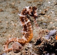
A thorny seahorse (Hippocampus histrix) - Classic case: a.k.a. “Hole in the head disease”
- Skin ulcerations
- Emaciation
- Dx:
- Etiology: Mycobacterium spp.
- Stress, crowding, poor water quality all make it worse
- Dx: necropsy shows internal granulomas (gray-white, necrotic foci), especially in liver, kidney, +/- spleen
- Dx: see acid-fast rods on cytology/histopath; PCR, culture
- Tx:
- None
- Bleach is ineffective at typical doses
- UV may help reduce loading, but mycobacteria thrive within biofilms and can be released
- Pearls: common!
- Zoonotic
- Syngnathids (seahorses) are very prone to infection; but likely mycobacteria can infect all fish species
- Risk factors: stress; excess organic debris, low pH, and low oxygen in the water
- Classic case: a.k.a. “Hole in the head disease”


Images courtesy of David Stanley (fish farm), Debbie Pouder (koi herpesvirus), Bernard Spragg (koi), Andreas R. Thomsen (louse), Robert Durborow (furunculosis), CDC/ Margaret M. Williams; Janice Haney Carr (Mycobacterium), and Nick Hobgood (thorny seahorse).
Top Topic Category
Exotics